Introduction
Medicine never ceases to amaze—and sometimes, reality sounds stranger than fiction.
In one such extraordinary case, a 33-year-old woman from Delhi faced an unimaginable condition: a portion of her brain tissue had slipped into her nasal cavity, causing months of severe nasal blockage and fluid discharge.
At HCMCT Manipal Hospital, Delhi, a team of expert neurosurgeons and ENT specialists performed an eight-hour life-saving surgery to restore her brain’s position and reconstruct the damaged skull base.
At The Doctorpreneur Academy, we highlight such groundbreaking cases that reflect the power of collaboration, innovation, and medical excellence—the very values driving the next generation of doctorpreneurs.
The Rare Case: When the Brain Breaches the Nose
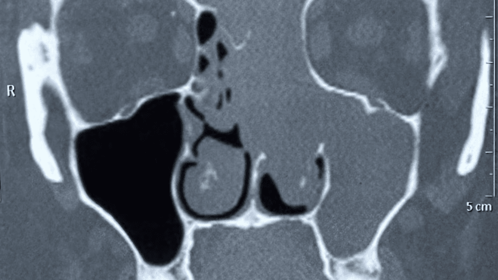
The patient’s condition was caused by a defect in the skull base, the thin bone that separates the brain from the nasal cavity. Over time, this bony partition eroded, allowing part of the brain to herniate downward — a rare condition medically known as encephalocele.
What made it alarming was that the brain tissue was visibly protruding into the nasal and oral cavities, posing a high risk of infection, cerebrospinal fluid (CSF) leakage, and even life-threatening meningitis.
She had suffered for months, visiting multiple clinics for nasal congestion and fluid discharge, but the true cause remained undiagnosed—until advanced imaging at Manipal revealed the shocking truth.
The Surgery: Precision, Teamwork, and Hope
Once diagnosed, doctors at HCMCT Manipal Hospital assembled a multidisciplinary team of neurosurgeons, ENT specialists, and anesthesiologists to plan a high-risk but necessary operation.
Key Details:
- Duration: 8 hours
- Approach: Combined endoscopic and transcranial surgery
- Objective: Reposition the herniated brain tissue and reconstruct the skull base
- Challenges:
- Risk of stroke due to proximity to major blood vessels.
- Potential for CSF leakage or infection.
- Maintaining brain integrity during repositioning.
Using state-of-the-art imaging and endoscopic technology, the team accessed the defect through the nose (minimally invasive route) and the skull (transcranial route).
They successfully lifted the displaced brain tissue back into its normal position, sealed the cranial defect, and reinforced the skull base with grafts—ensuring both structural stability and safety.
The Outcome: A New Lease on Life
Post-surgery, the patient showed remarkable recovery. Her nasal symptoms subsided, brain function remained intact, and she was discharged in stable condition.
She continues to be under follow-up care, living a normal, healthy life once again.
💬 “It was one of the rarest and most delicate surgeries we’ve performed,” said one of the operating surgeons. “Every minute counted—but teamwork made it possible.”
The Doctorpreneur Perspective: Lessons from This Case
This case underscores the future of multidisciplinary healthcare, where teamwork and technology converge to save lives.
Here are a few takeaways for modern doctors and innovators:
✅ 1. Collaboration is the new clinical power.
Complex cases require cross-specialty partnerships—ENT, neurosurgery, radiology, and anesthesiology all worked in sync here.
✅ 2. Technology is the surgeon’s strongest ally.
From endoscopic cameras to high-resolution imaging, precision tools enabled minimal damage and faster recovery.
✅ 3. Early diagnosis saves lives.
Persistent nasal discharge or blockage can indicate deeper issues. Empowering general physicians and ENT doctors to spot early red flags is crucial.
✅ 4. Patient education and access matter.
If patients understand warning signs, they seek help sooner—reducing late-stage complications.
✅ 5. The future is in innovation and compassion.
This case represents the kind of innovation that Doctorpreneur Academy encourages—where technology, empathy, and skill unite for better outcomes.
What It Means for Doctors in India
For Indian healthcare professionals, this case is a reminder of the immense potential of modern neurosurgery and interdisciplinary medicine.
It also highlights how infrastructure, innovation, and trained specialists can bring global standards of care to Indian hospitals.
As doctorpreneurs, doctors can:
- Advocate for advanced diagnostics in ENT and neurology.
- Participate in interdisciplinary simulation and surgical workshops.
- Support public awareness initiatives about rare cranial conditions.
When doctors innovate and communicate better, patients benefit sooner—and lives are saved.
Conclusion
A brain herniating through the nose sounds like a plot from a medical thriller—but for one woman, it was reality.
Thanks to the skill, precision, and teamwork of doctors at HCMCT Manipal Hospital, she not only survived but also regained a normal life.
This case exemplifies what modern medicine and collaborative innovation can achieve.
In a remarkable medical achievement, doctors at HCMCT Manipal Hospital, New Delhi, successfully treated a 33-year-old woman whose brain tissue had herniated into her nasal cavity due to erosion of the cranial base—a condition known as encephalocele. The patient had been suffering for months from nasal blockage and fluid discharge before undergoing a life-saving eight-hour surgery. Using a combination of endoscopic and transcranial techniques, the multidisciplinary team carefully repositioned the brain tissue and reconstructed the skull base.
The surgery, which carried high risks of stroke and major blood vessel damage, was completed successfully, and the patient has now fully recovered and resumed normal life.
👉 To register for our next masterclass, please click here: https://linktr.ee/docpreneur